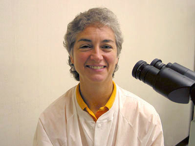
| Authors | ||||||||||||
 |
Linda M.
Marler, M.S., MT(ASCP)SM Associate Professor Education Coordinator, Division of Clinical Microbiology Department of Pathology and Laboratory Medicine Indiana University School of Medicine | |||||||||||
 |
Jean A.
Siders, M.S., MT(ASCP) Assistant Professor Director, Pathology Multimedia Education Group Department of Pathology and Laboratory Medicine Indiana University School of Medicine | |||||||||||
 |
Cynthia Kaufman, M.S., MT(ASCP)SM Technical Coordinator Division of Clinical Microbiology Clarian Health Partners (Methodist-IU-Riley) Indianapolis, IN |
|||||||||||
 |
Erick Owens, MLT (ASCP) Division of Clinical Microbiology Wishard Hospital Indianapolis, IN |
|||||||||||
 |
Greg Korba , MT (ASCP) Division of Clinical Microbiology Clarian Health Partners (Methodist-IU-Riley) Indianapolis, IN |
|||||||||||
 |
Stephen D.
Allen, M.D. Professor Department of Pathology and Laboratory Medicine Indiana University School of Medicine Director, Division of Clinical Microbiology Clarian Health Partners (Methodist-IU-Riley) | |||||||||||
|
Ryan Christy, B.S. Brent Gann, B.A. Shadi Zakhour, B.F.A.
Special Kudos : Special thanks to Caroyln Biggs for her constant support and for her editorial expertise in reviewing this CD. Our thanks to Jim Cassell for his support and photographic expertise that was used for the CD cover.
The Indiana Pathology Image Atlas Series™ Bacteriology I CD, containing nearly 700 quality photomicrographs, was developed for the purpose of providing a visual atlas that would serve as a valuable resource for educators, students and practitioners. Space restrictions necessitated the separation of the vast scope of material into more than one Bacteriology Image Atlas. The first in this series, Bacteriology I, contains many of the more commonly encountered organisms in clinical microbiology and a few less commonly encountered organisms of educational interest. Images in the CD can be accessed using one of the following tabs: “Alphabetical”, “Grouping”, or “Tests”. The authors recognize that some bacteria could be included in more than one group; however, the following arbitrary groups were used: “aerobes”, e.g. obligate aerobes, facultative anaerobes and microaerophilic organisms and “anaerobes” e.g. obligate anaerobes, aerotolerant anaerobes and the occasional facultative and microaerophilic organisms. Full plate, close-up and/or stereoscopic views are included for many isolates. The importance of stereoscopic examination of anaerobic organisms can’t be over-emphasized; hence, the authors chose this magnification as the opening image for viewing all anaerobe isolates. All isolates were incubated until colony morphology was well established; 24 hours for most fermenting “aerobes” and 48 hours for most non-fermenting “aerobes” and “anaerobes”. Since this CD has been designed for use as a “visual” review and image resource, text has been limited to essential information. For detailed discussion and descriptions, the authors recommend use of other clinical microbiology resources (e.g., textbooks, websites).
Image Atlas Series The Indiana Pathology Images™ Image Atlas Series is a CD project developed for educators, students, and practitioners. This series offers affordably priced CDs, each containing a comprehensive collection of clinically relevant images in a "click-art" format. Images are high quality with high resolution for viewing and printing purposes, and appropriately sized for easy use in most software programs. Images have been digitally edited only to improve image quality. Each CD in the series is a comprehensive atlas of images that will eliminate the need to scan pictures and slides or search the internet for teaching materials. Each title is an invaluable educational resource and certain to enhance any image library. Suggested uses for Indiana Pathology Images™
products
inQUIZator Series Indiana Pathology Images™ developed the inQUIZator™ series in answer to requests for affordable, quality review and testing materials in areas of pathology and laboratory medicine. These CDs are composed of questions, half of which include high-quality images. inQUIZator CDs offer versatility with user options including review, self-test and competency test modules. The review module allows the user to select a test bank sorted by chapters, e.g. lab procedures, identification, disease, etc., or by alphabetical listing. The review module also offers a randomized self-test. The competency test module allows the user to take a randomized test selected from a bank of questions with images that will assess the user’s ability to identify organisms. After completion of the self-test or competency test, a certificate can be printed that includes the user’s name, date and test results. This certificate can be used to document competency for laboratory personnel, students, residents and fellows.
Visit our web site at www.ipimages.com for a list of current and future Image Atlas and inQUIZator™ CD titles. Copyright © 2005 Indiana Pathology Images™ All rights reserved. Images on this CD are protected by copyright. No image may be reproduced in any form or by any means and used for publication without the expressed written permission from the copyright owner. The publisher is not responsible (as a matter of product liability, negligence, or otherwise) for any injury resulting from any material contained herein.
Minimum System Requirements
Installation Procedure (recommended for optimum operation)
Run from CD-ROM
CD Features
Sales Technical Support Comments or Questions Visit Our Website
| ||||||||||||